I've been working on my next post, but won't be able to finish it until after the weekend, as I have to attend a wedding and participate in copious amounts of "family stuff".
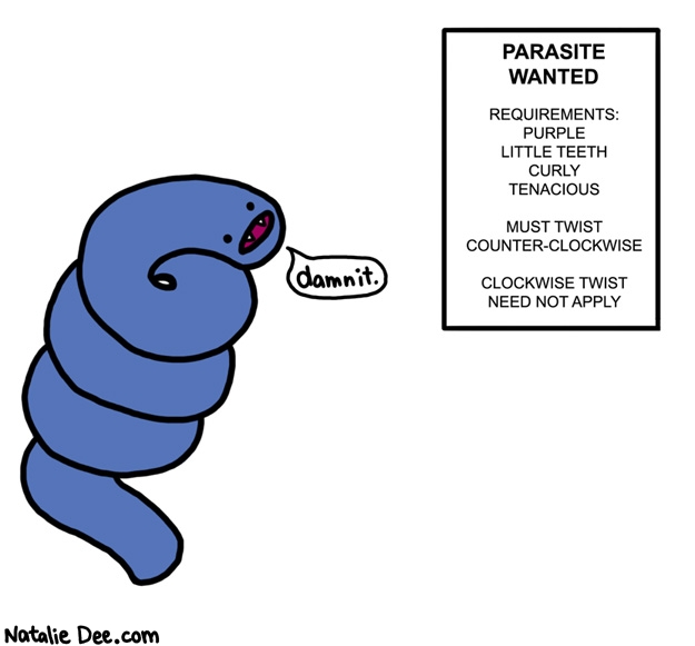
Until then, I'll leave you with the awesome new post from Natalie Dee:
I'm sure I'm the one that found that funny. I'll shut up now.
I know, I know, I touched on malaria in my last entry. But, World Malaria Day is tomorrow, April 25th.
Malaria is one of the most prevalent and debilitating diseases that is currently affecting our species. A lot of scientific advancements have come into fruition because our struggle with battling malaria. Since its World Malaria Day, I’ll tell you a little bit about the background and history of malaria.
Most people assume malaria is a disease that is only concentrated in Africa or some tropical regions of Asia, but that is not the case. In 1937, there were over 1 million cases of malaria in the United States, and it currently affects people in hundreds of countries. Malaria cases were eradicated from the United States over 50 years ago, but more than 40% of the world’s population is at risk. Although over 90 percent of malaria cases today are in sub-Saharan Africa, it affects the entire world.
Many people don’t give malaria the credit it deserves for being such a horribly intelligent and sneaky parasite. Some historical epidemiologists believe that malaria played an important hand in the fall of ancient Greek and Roman empires. In medieval times, many expeditions were not wiped out by the wars they were engaged, but by malaria infections instead. During the Vietnam war, battle wounds were the most prevalent reason for hospitalization; Malaria was the second.
In 1955, the World Health Organization proposed to eradicate malaria worldwide through the use of dichlorodiphenyltrichloroethane, or DDT. Their efforts began with routine house spraying, and “bombing” areas of high infectivity or environments that were conducive to mosquito breeding. The emergence of drug resistance, widespread resistance to available insecticides, wars and massive population movements, difficulties in obtaining sustained funding from donor countries, and lack of community participation made the long-term maintenance of the effort untenable. Completion of the eradication campaign was eventually abandoned to one of control.
According to the Malaria No More organization, malaria kills an African child every 30 seconds. In 2008, an estimated 863,000 people died of malaria Although these facts and statistics may make it seem so, a malaria infection and diagnosis is not a death sentence. Malaria is definitely a treatable and curable disease.
Twitter has joined up with Malaria No More and other organizations for World Malaria Day and has set up a program where you can donate $10 with every retweet you send out. Check it out here.
Also note, the World Health Organization just announced on April 23rd:
Rapid diagnostic malaria tests will help health workers quickly identify which patients have the disease and need immediate treatment. This will allow malaria to be properly diagnosed before treatment.
I encourage you to take a moment and think about how you have screens on your windows, access to anti-malarial drugs, and don’t need to sleep under a net to ensure your survival.
I didn’t think it would happen, but I actually had a request for my next blog entry!
I can’t exactly tie this one into any overly religious, candy-laden holiday, but it happens to be one of my more favored parasites that are often mistaken for a virus… any guesses?
Malaria!
I’m not sure why, but I’ve heard many people refer to malaria as a viral infection, when it is actually a hemolytic parasite. Maybe it’s the fact that early malaria was associated with the air content of the prevalent environments (often murky or musty), which gave rise to the naming of malaria-- meaning “bad air”. This can be drawn to some viruses and their airborne, or aerosolized, transmission technique. Yet, we (as in…scientists, duh.) quickly discovered that malarial transmission was not through aerosolization of a virus, or from any direct contact. The only other similarity that the parasite genus Plasmodium has to common viral infections is the series of flu-like symptoms, such as fever and chills. In initial diagnosis trials, malaria was often misdiagnosed as an ongoing influenza with rhythmic symptom cycles. Seasonal influenza causes anywhere between 250,000 and 500,000 deaths annually, whereas Malaria causes approximately 1 million deaths annually. Needless to say, there are major differences.
So, now that we know Malaria is, in fact, a parasite, the details of its lifecycle can help answer many further questions (also, one specifically that has been requested).
Malaria is actually, roughly, an umbrella term that can be used to describe the disease in humans that is caused by four parasites: Plasmodium vivax, Plasmodium falciparum, Plasmodium malariae and Plasmodium ovale.
Plasmodium ovale is regionally specific to West/sub-Saharan Africa, with a much lower prevalence (~5%) in places like the Philippines, Papua New Guinea and Cambodia. P. ovale is very specific in region and may seem to have a stippled appearance. P. ovale is also the only form of malaria that is fibronated.
Plasmodium ovale: trophozoite after "ring stage"
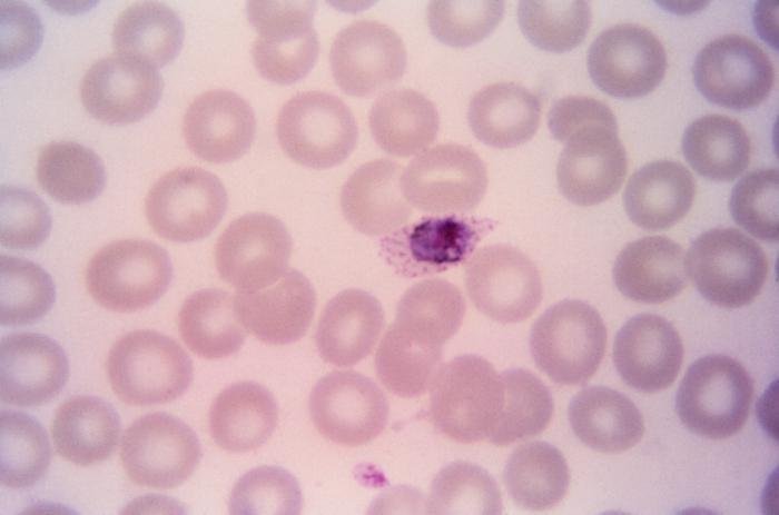
Plasmodium vivax is the most common form of malaria that causes reoccurring symptoms, or is tertiant in its cycle. P. vivax is found widely spread over Asia, Latin America and some parts of African, and causes debilitating, reoccurring symptoms that are mostly non-fatal. It can also cause splenomegaly, or enlargement of the spleen, which is highly fatal.
Plasmodium vivax: immature schizonts in human blood smear
Plasmodium malariae is very similar to P. ovale, in that it causes “milder”, or less fatal symptoms in very specific areas. P. malariae is thought to be the oldest form of Malaria causing parasites, and is relatively non-commensal. P. malariae undergoes a quartant cycle, replicating every 72 hours. Sometimes, the meroxoites form in a rosette fashion. My parasitology professor always used to say “if you have to choose which type of malaria to be infected with, choose Plasmodium malariae.”
…right. It’s so convenient how those Anopheles are giving you a choice, these days.
Plasmodium malariae: trophozoite illustrating the "ring stage" in human blood smear
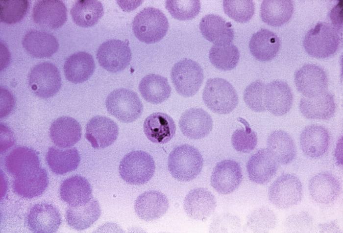
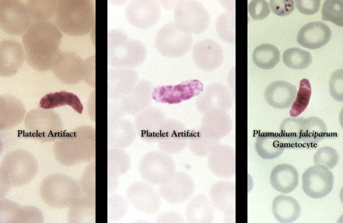
Now, my favorite, Plasmodium falciparum is the most deadly of the four. To illustrate its fatal nature, it is shaped like a banana. Yea, a deadly banana. Aren’t you scared?
P. falciparum is often termed as the “newest” of the four because it is so dangerous and deadly. In recent years, it has accounted for more than 95% of all malarial infections, and over 90% of all the deaths caused by malarial infections. P. falciparum has the unique ability to infect one cell with multiple merozoites. Essentially, P. falciparum is like a sniper version of malaria: it gets the job done. Like I said, deadly banana.
Plasmodium falciparum: banana shaped gametocytes in human blood smear
The lifecycle of malaria occurs in two parts: the sexual cycle that occurs within the vector’s gut, and the asexual cycle that occurs within the human host’s tissues and circulatory system.
Within the human, the Plasmodium sporozoites infect the liver cells and release merozoites. These merozoites infect red blood cells, replicate, and cause the red blood cells to literally rupture and explode in order to infect other red blood cells nearby. Eventually, this causes chaos in your liver tissues and circulatory system.
Yet, interestingly enough, if you already have Thalassemia, you have the added bonus of an evolutionary “immunity” to malaria diseases. Thalassemia is a disease associated with globin gene mutations (both α- and β-globin genes can be mutated, yet α-globin mutations are most common). Since your α- or β-globin gene is recessively mutated if you have active Thalassemia, your red blood cells develop into malformed, or sickled, cells, thus inhibiting their affinity to bind and transport oxygen. This lower affinity can cause extreme cases of anemia. In major cases, treatments such as chronic blood transfusion therapy, splenectomy, transplantation and iron supplementation are used.
Many immunologists believe that this immunity to malaria diseases that is associated with Thalassemia is a result of Darwinian genetics. A good example of this is Africa, which has some of the highest rates of malaria in the world, as well as a Thalassemia diagnosis of approximately 40% of the entire population. Those with the recessive mutation survive malaria more effectively, and have a higher likelihood of handing such mutations down through to following generations.
On a semi-related note, a friend turned me on to this Animal Planet show called Monsters Inside Me, which is all about parasites and other fun things! Here is a short video about malaria with some neat animations of the active infection:
I’m sure I will go into more specifics about malaria in future posts, but I wanted to address the question and request that I received in a timely manner. With that being said, if you have a topic that you are dying to read about, please feel free to send me an email, and I’ll get to it!
I had a difficult time deciding what to kick off my first blog entry with – there are so many viruses and parasites to choose from! Do I start with something basic and move onwards and upwards from there? Or do I jump right in with both feet and bombard my initial readers with my favorites?
But then I realized I should obviously go with a festive theme for Easter. I mean, that’s the logical thought, right?
Truth be told, the Easter bunny does become occasionally ill. Unfortunately, it’s usually with a brutally fatal virus: the myxoma virus.
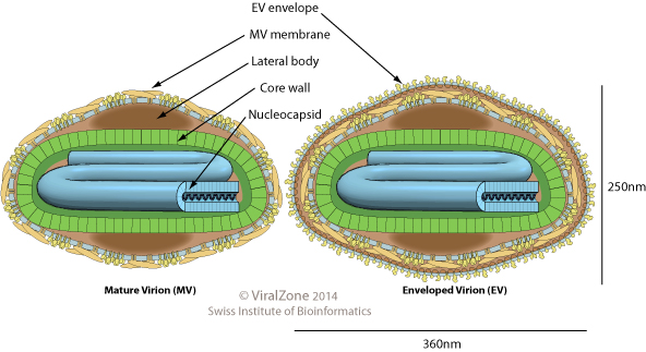
The myxoma virus is part of the poxviridae family, and is a class 1 virus within Baltimore Classification Standards. The myxomoa virus has an exterior envelope structure which encases the viral capsid and assists with viral entry to the host’s cell. This envelope structure contains many important glycoproteins that assist with entry through membrane fusion and location specific phagocytosis. Later on in the viral lifecycle, or the “infectivity cycle”, the viral envelope also assists with budding from the host’s cell for dissemination of newly matured virions. For all viruses, the viral genome is protected within the viral capsid. The myxoma virus utilizes a double stranded DNA genome that is non-segmented and linear.
The myxoma virus only seems to infect rabbits and hares due to components of their immune system that are easy to evade. It is not necessarily exclusive to one geographic region, although does have a higher infectivity in specific areas. High incidence rates have been reported in the UK, Australia, Egypt, France and South American (specifically Uruguay). The South American incidents are typically not dangerous and illustrate a lower mortality rate than any other region affected. Common cottontail rabbits usually experience characteristic dermal tumors, yet European rabbits can experience severe myxomatosis.
Symptoms of myxomatosis begin with tumors on the skin and genital areas. After spreading, the rabbits can experience blindness, fever and exhaustion. Due to the rabbit’s weakened state, secondary bacterial infections are very common and are habitually what cause the onset of the eventual death. This is usually due to swelling of the lung tissues and pneumonia. Myxomatosis is also associated with Rabbit Hemorrhagic Disease (RHD) which can cause the infected rabbit to become comatose and die. Death from a myxoma infection can occur anywhere between 48 hours to 14 days after initial infection.
Australia’s government decided to utilize the fatality of the myxoma virus to control the overwhelming rabbit population. Initial experiments for population control “successfully” reduced the rabbit population from 600 million rabbits to 100 million rabbits over the course of two years. I can’t imagine what wading through rabbit carcasses decorated with tumors would be like. I mean, 500 million is a lot of dead rabbits.
The virus is spread through direct contact with infected rabbits, and also through insects that take blood meals, such as mosquitoes. It’s still a pretty rampant problem, thanks to governmental abuse for population control, and has lead to the development of a vaccine for those who wish to keep pet rabbits.
So, don’t be alarmed if the Easter Bunny didn’t quite make it through his rounds of delivering baskets. You might be the lucky one that finds a lumpy rabbit carcass amongst your chocolate eggs.
As far as festive parasitic infections go, Iodamoeba butschlii takes the egg-shaped cake.
Iodamoeba butschlii, as you can see from the name, is an amoebic parasite. It does infect humans, but is considered mostly non-pathogenic. Generally, these little buddies reside in your large intestines and feast on the excess yeast in your system. Rarely do people experience symptoms, but if they do, it’s generally just mild diarrhea and stomach cramps.
The festivity is in the structure of the parasite, not necessarily the function (I mean, nothing says Happy Easter like mild diarrhea and stomach cramps!). Iodamoeba butschlii is one of the smaller parasites, with an average cyst size of 10 µm. It has all the normal components of an amoebic parasite, such as a nucleus, endosomes, etc., yet can be easily diagnosed because of its abnormally large glycogen vacuole! Why am I so excited about this glycogen vacuole, you wonder? Well, it is the key to our festive riddle!
Scientists, being the witty and intelligent creatures that they are, decided that this glycogen vacuole paired with the ovoid shape of the cyst makes Iodamoeba butschlii look like an EASTER BASKET! Take a look for yourself:
Don't you just want to put all your eggs in that little basket? An Iodamoeba butschlii infection can be diagnosed through a fecal wet mount. Exciting!
So with that, watch out for excess rabbit carcasses and avoid eating focally contaminated food this Easter.


